 заплесневелый
Ядерный Магнитно-резонансная томография (NMRI, Nuclear Magnetic Resonance Imaging), also known as Magnetic Resonance Imaging (МРТ, Магнитно-резонансная томография), is a kind of nuclear physical phenomenon. The physical basis of MRI is the theory of Nuclear Magnetic Resonance (ЯМР) The so-called NMR refers to the resonance phenomenon related to the magnetic and magnetic fields of a substance. It can also be said that it is a low-energy electromagnetic wave, то есть, the physical characteristics exhibited by the interaction between the radio frequency wave and the nuclear system with both angular momentum and magnetic moment in the external magnetic field. Using this phenomenon can not only study the composition of the substance, but also observe its microstructure.
According to this, people use various radio frequency pulse sequences to stimulate organisms, and use detection coils to record tissue relaxation, proton density, flow, chemical shift, диффузия, perfusion, blood oxygenation status, and tissue temperature. МРТ-технология. From the discovery of NMR to the birth of MRI device, there have been decades of hard work by several generations of physicists and medical scientists.
As early as the 1930s, the physicist Isido Isaac Rabbi discovered that the nuclei in the magnetic field would be arranged in parallel in the order of forward or reverse along the direction of the magnetic field. The direction of rotation is reversed. This is the earliest human understanding of the interaction of atomic nuclei with magnetic fields and applied radio frequency fields. В 1946, American physicists Felix Bloch and Edward Purcell discovered that the nuclei in the magnetic field would be tilted when excited by high-frequency electromagnetic fields. When the high-frequency field is turned off, the nucleus will release the absorbed energy and return to the original state process). From this, the phenomenon of Time-domain nuclear magnetic resonance (ТД-ЯМР) was discovered, and earlier it was only used in chemical analysis. Because of his outstanding contribution to the theoretical basis of nuclear magnetic resonance imaging, Isido Isaac Rabbi won the Nobel Prize in Physics in 1944, and Felix Bloch and Edward Mills Persell shared Nobel Prize in Physics in 1952.
В 1950, Owen Hahn discovered the phenomenon of nuclear magnetic resonance spin echo under double pulse. At the beginning of the discovery of the nuclear magnetic resonance phenomenon, due to the defects of harsh imaging conditions and long imaging time, the scope of application was greatly limited. Until 1968, the team of Richard Ernst improved the excitation pulse sequence and analysis algorithm to greatly improve the signal. After the sensitivity and imaging speed, the nuclear magnetic resonance technology gradually matured, and Richard Ernst himself won the Nobel Prize in Chemistry in 1991.
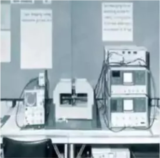
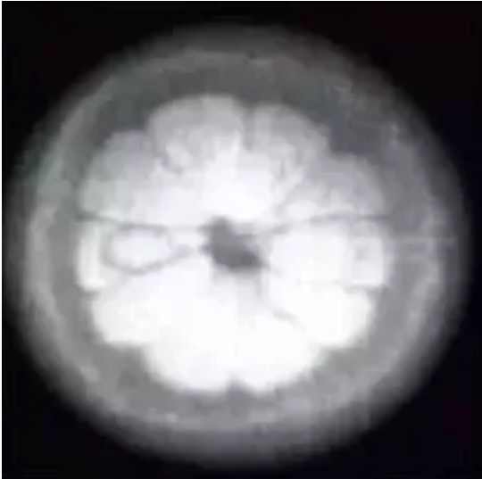
В 1973, American chemist Paul Lauterbur (University of Illinois at Urbana, was born on May 6, 1922, Ohio, США) and British physicist Peter Mansfield School of Astronomy, born on October 9, 1933 in London, England) completed the initial nuclear magnetic resonance imaging system (Фигура 1) in the central laboratory of the Netherlands, and imaged liquid-filled objects to obtain the famous nuclear magnetic resonance image “Nottingham’s Oranges” (Фигура 2) won the top spot in the field of nuclear magnetic resonance imaging. Making it into the field of medical imaging has become one of the most important imaging methods today.
The 2003 Nobel Medical Prize jury announced at the Karolinska Institute of Medicine in Sweden that the Nobel Prize in Physiology or Medicine was awarded to two scientists, Paul Lauterbur and Peter Mansfield, in recognition of their nuclear magnetic resonance imaging Outstanding contribution made by technology. The former invented the gradient field, which gradually matured the nuclear magnetic resonance imaging technology; the latter conducted pioneering research on the imaging method and invented the echo planar imaging technology, which made the image quality better and the imaging process faster. MRI is another medical imaging achievement that has won the Nobel Prize after the discovery of X-rays (the Nobel Prize in Physics in 1901) and the invention of X-ray CT (the Nobel Prize in Physiology or Medicine in 1979). The highest award in the scientific community fully affirmed the great significance of MRI in human medical and health care activities, and also greatly inspired scholars in this field.
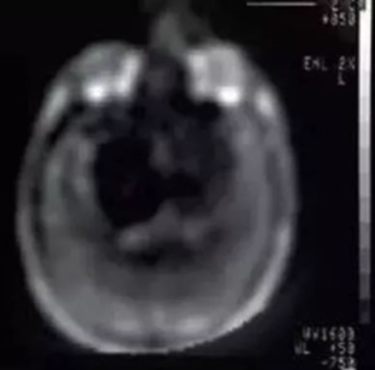
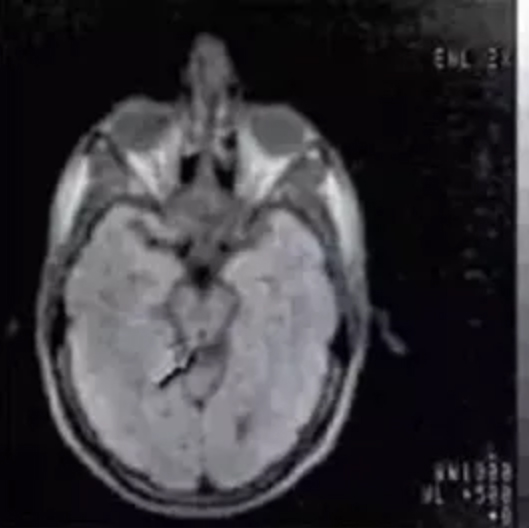
Encouraged by the imaging results, в 1978, the Dutch Central Laboratory formed a “proton project” research team, which developed a 0.15T nuclear magnetic resonance system, and on December 3, 1980, obtained the first human head MRI image (Фигура 3) and the first image after the second-dimensional Fourier transform (Июль 30, 1981, Фигура 4).
Всего за 50 годы, nuclear magnetic resonance imaging technology has made great progress and has become one of the four routine inspection methods of imaging (four conventional methods: ядерно-магнитно-резонансная томография, X-ray imaging, ultrasound imaging and nuclear medicine imaging). В отличие, nuclear magnetic resonance imaging has a high resolution of soft tissues, and the advantage of no radiation damage makes it irreplaceable in infant development and bone ligament strain. The journey that MRI has taken from physical research to chemical and biological applications to clinical applications is the most convincing example of the development of basic science today that promotes social progress. With the advancement of superconducting technology, cryogenic technology, magnet technology, electronic technology, imaging technology, image processing technology and computer science technology, MRI equipment and imaging methods have achieved rapid development. The advancement of technology has continuously expanded its application fields.
In the direction of development, nuclear magnetic resonance systems are constantly pursuing extreme working conditions and more targeted excitation sequences. In the field of nuclear magnetic resonance imaging, there are two development trends, one is the direction of superconductivity, also known as HF-NMR, and the field strength is generally above 1.5T; the other is the direction of permanent magnetism, also known as TD-NMR, and the field strength is 0.2 Between T-0.35Т.