Small Animal Imaging by NMR Instrument
Small Animal Imaging Application
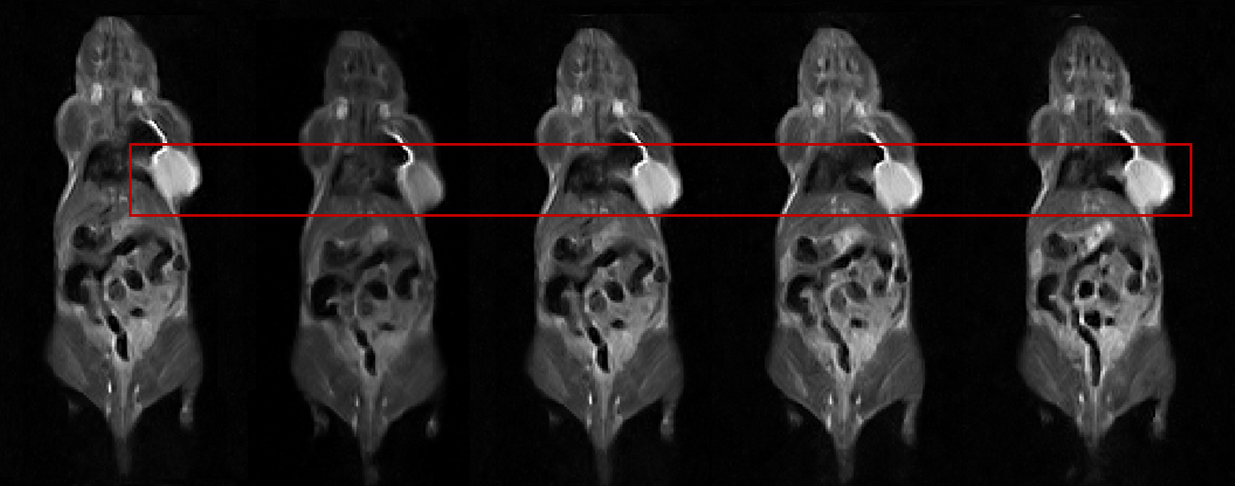
Small animal imaging plays a crucial role in various aspects of biomedical research, including targeted agent contrast agent studies, anatomy teaching, and biomedical clinical animal experiments. The ability to perform in vivo imaging of small animals such as mice, rabbits, and monkeys allows researchers to gain a deeper understanding of their bodies and observe subtle differences between tissues and organs. Magnetic Resonance Imaging (MRI) is a commonly used technique for small animal imaging due to its ability to provide detailed and non-invasive images.
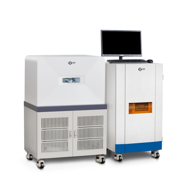
Small Animal Imaging Analyzer of Niumag
Niumag, a leading provider of small animal MRI technology, has extensive experience in the field of low-field nuclear magnetic resonance. They have developed advanced systems specifically designed for small animal imaging, offering a range of different caliber options from 15mm to 150mm. These systems incorporate contrast agent relaxation analysis capabilities, enabling researchers to perform targeted observations and research on small animal subjects.
Small animal imaging tests using MRI offer several valuable applications:
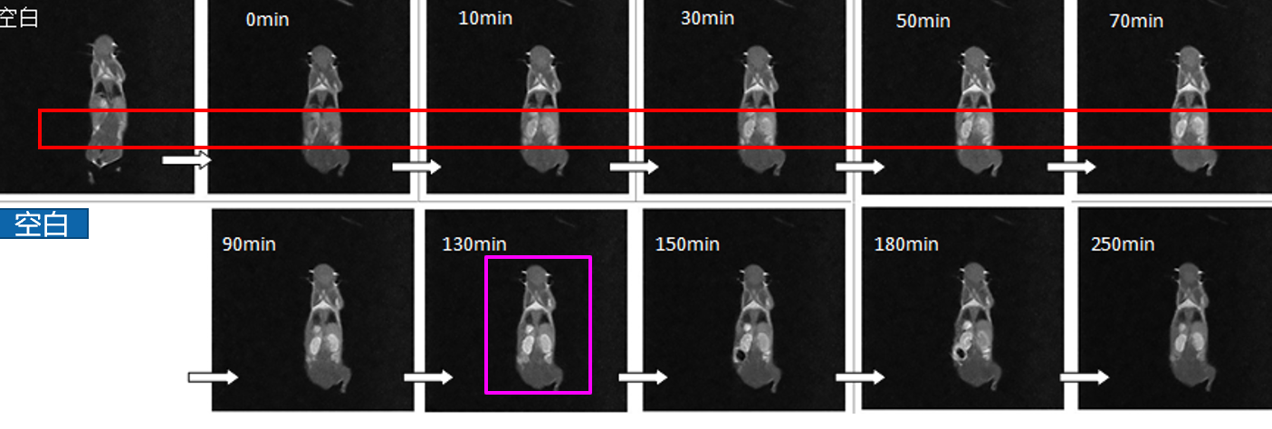
- Evaluation of the effect of contrast agents in vivo: By administering contrast agents and conducting MRI scans, researchers can assess how these agents interact with tissues and organs in living animals. This evaluation helps in understanding the dynamics and effectiveness of various contrast agents.
- Determination of drug targeting: Small animal imaging allows researchers to track the distribution and accumulation of drugs within the body. By visualizing the targeting behavior of drugs, researchers can optimize drug delivery strategies and enhance therapeutic outcomes.
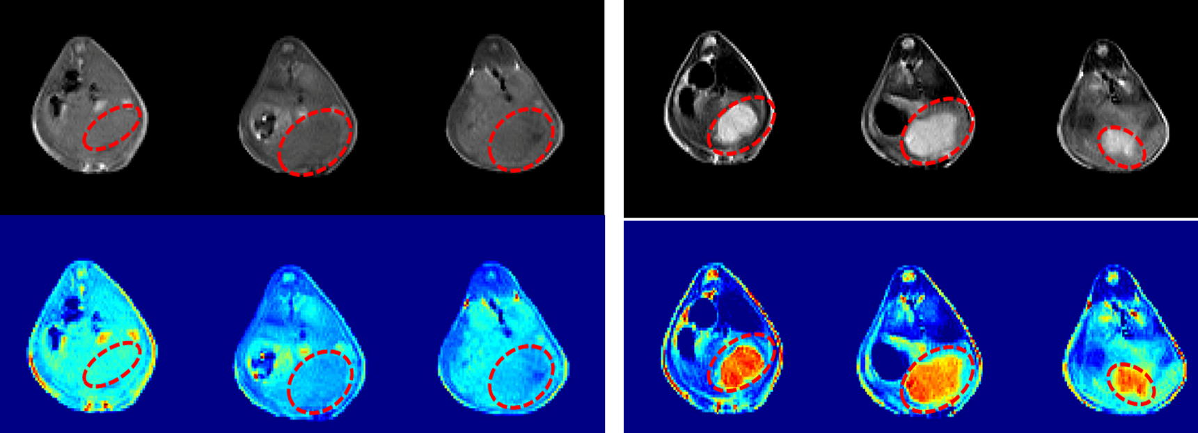
- Evaluation of the therapeutic effect of drugs on tumors: MRI enables the monitoring of tumor growth, response to treatment, and assessment of therapeutic outcomes. By comparing pre- and post-treatment scans, researchers can evaluate the effectiveness of drugs in inhibiting or reducing tumor size.
- Location of tumor lesions: Small animal MRI facilitates precise localization of tumor lesions, aiding in the accurate characterization and diagnosis of tumors. This information is essential for planning interventions, surgeries, or further targeted studies.
Additionally, MRI evaluation of rat kidney size provides valuable insights into renal morphology and function. By visualizing the kidneys through MRI, researchers can accurately measure their dimensions and assess any structural abnormalities. This information contributes to a better understanding of kidney health and the effects of various experimental conditions or treatments on renal structures.
Furthermore, MRI analysis of the whole-body composition of rats allows researchers to investigate and quantify the distribution of different tissues within the animal’s body. This analysis aids in understanding changes in body composition due to factors such as diet, disease, or drug interventions. By segmenting and analyzing MRI images, researchers can determine the relative proportions of muscle, fat, and other tissues, providing valuable data for physiological and preclinical studies.
Small animal imaging, particularly through MRI, is an invaluable tool in biomedical research. It enables researchers to non-invasively visualize and analyze small animal subjects, contributing to advancements in drug development, disease understanding, and therapeutic interventions. Niumag’s expertise in small animal MRI technology further enhances the capabilities and precision of these imaging studies.
 NIUMAG


WeChat
Scan the QR Code with WeChat