Визуализация мелких животных by NMR Instrument
Визуализация мелких животных Приложение
Визуализация мелких животных plays a crucial role in various aspects of biomedical research, including targeted agent contrast agent studies, преподавание анатомии, and biomedical clinical animal experiments. The ability to perform in vivo imaging of small animals such as mice, кролики, and monkeys allows researchers to gain a deeper understanding of their bodies and observe subtle differences between tissues and organs. Магнитно-резонансная томография (МРТ) is a commonly used technique for визуализация мелких животных due to its ability to provide detailed and non-invasive images.
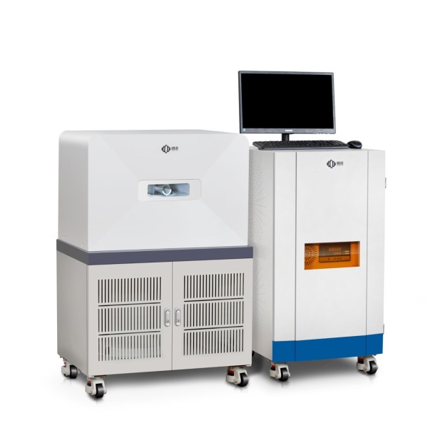
Визуализация мелких животных Analyzer of Niumag
ответил, ведущий поставщик технологий МРТ для мелких животных, имеет большой опыт работы в области низкополевого ядерного магнитного резонанса.. They have developed advanced systems specifically designed for визуализация мелких животных, предлагает широкий выбор вариантов калибра от 15 мм до 150 мм.. Эти системы включают возможности анализа релаксации контрастного вещества., enabling researchers to perform targeted observations and research on small animal subjects.
Визуализация мелких животных tests using MRI offer several valuable applications:
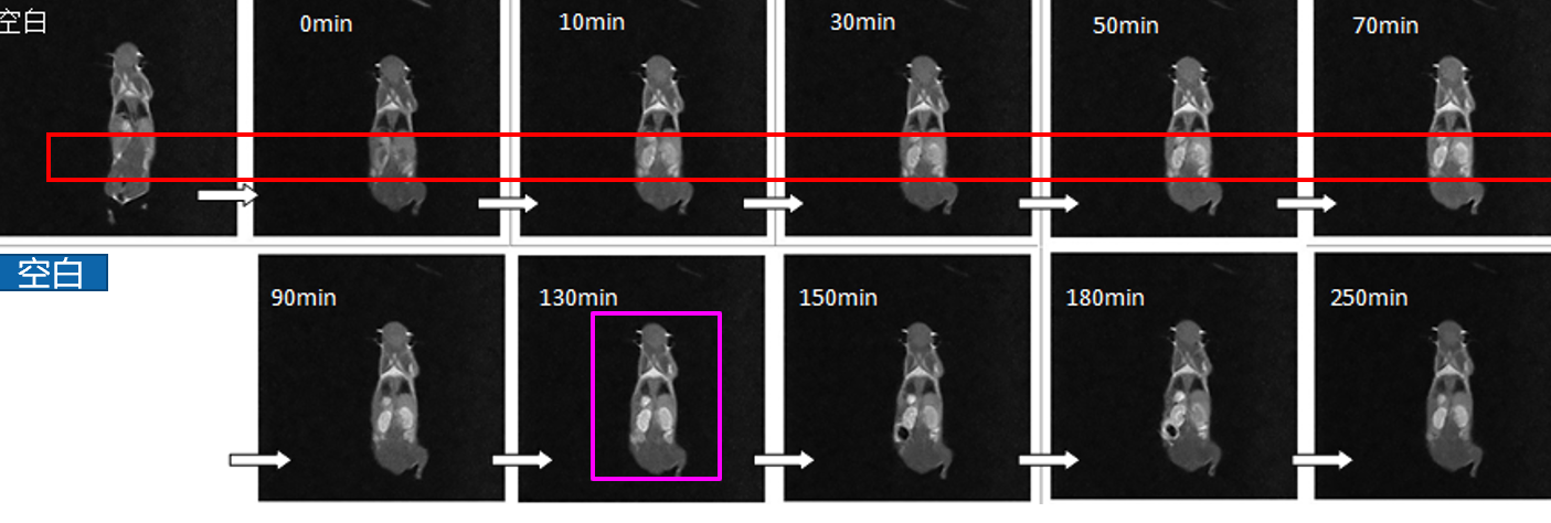
- Оценка эффекта контрастных веществ in vivo: By administering contrast agents and conducting MRI scans, researchers can assess how these agents interact with tissues and organs in living animals. This evaluation helps in understanding the dynamics and effectiveness of various contrast agents.
- Определение таргетной направленности препарата: Визуализация мелких животных allows researchers to track the distribution and accumulation of drugs within the body. By visualizing the targeting behavior of drugs, researchers can optimize drug delivery strategies and enhance therapeutic outcomes.
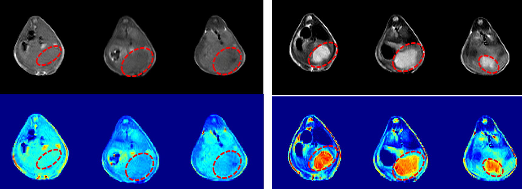
- Оценка терапевтического действия препаратов на опухоли: MRI enables the monitoring of tumor growth, response to treatment, and assessment of therapeutic outcomes. By comparing pre- and post-treatment scans, researchers can evaluate the effectiveness of drugs in inhibiting or reducing tumor size.
- Расположение опухолевых поражений: Small animal MRI facilitates precise localization of tumor lesions, aiding in the accurate characterization and diagnosis of tumors. This information is essential for planning interventions, surgeries, or further targeted studies.
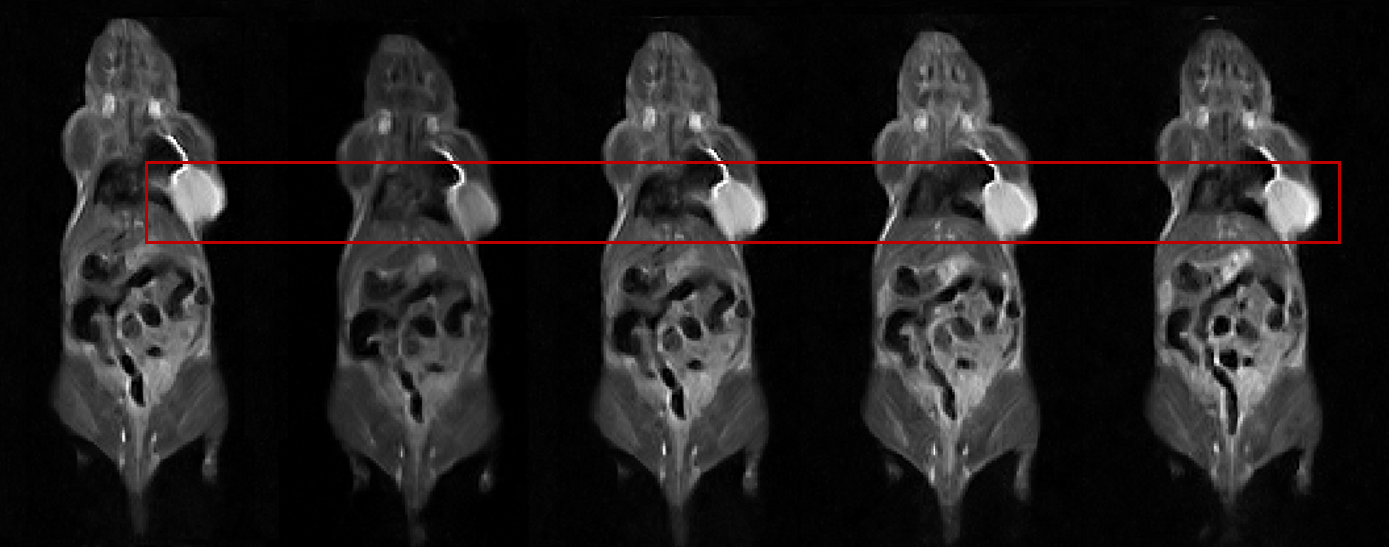
Кроме того, MRI evaluation of rat kidney size provides valuable insights into renal morphology and function. By visualizing the kidneys through MRI, researchers can accurately measure their dimensions and assess any structural abnormalities. This information contributes to a better understanding of kidney health and the effects of various experimental conditions or treatments on renal structures.
Более того, MRI analysis of the whole-body composition of rats allows researchers to investigate and quantify the distribution of different tissues within the animal’s body. This analysis aids in understanding changes in body composition due to factors such as diet, disease, or drug interventions. By segmenting and analyzing MRI images, researchers can determine the relative proportions of muscle, толстый, and other tissues, providing valuable data for physiological and preclinical studies.
Сmall animal imaging, particularly through MRI, is an invaluable tool in biomedical research. It enables researchers to non-invasively visualize and analyze small animal subjects, contributing to advancements in drug development, disease understanding, and therapeutic interventions. Niumag’s expertise in small animal MRI technology further enhances the capabilities and precision of these imaging studies.
 заплесневелый


