Система МРТ для мелких животных в оценке черепно-мозговой травмы у крыс
МРТ мелких животных-мощный неинвазивный инструмент, который можно использовать для обнаружения широкого спектра поражений.
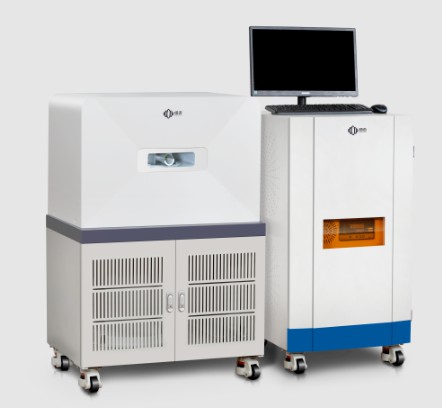
Компания Niumag разработала и выпустила новый компактный, высокопроизводительная система МРТ для мелких животных, в которой используется конструкция ЯМР с низким полем. Эта система МРТ с небольшими животными является основанным на приложении подхода, снизить стоимость и сложность традиционных систем. Система МРТ с мелким животным мобильной и самоочищенной и может быть размещена в большинстве исследовательских объектов. От этого мелкого животного не требуется запас хладагента или коммунального предприятия. Преимущество этой системы МРТ с мелким животным состоит в том, что она может легко обеспечить четкие 3D цифровые морфологические изображения всего органа целевого органа.
В анализе МРТ мелких животных, Время релаксации спин-латши (Т1) и время релаксации спин (Т2) Значения часто встречаются, нормальные закономерности которых варьируются в зависимости от органа, салфетка, и жидкость . Когда травма головного мозга была вызвана у крыс, Сигналы T1 и T2 были соответственно изменены из -за изменений ткани; Эти изменения изображения T1 и T2 позволили нам обнаружить индуцированную травму головного мозга.
Пилокарпин, мускариновый холинергический агонист, широко распространенный препарат для индукции эпилепсии и морфологического повреждения нейронов в мозге крысы. В этой модели травмы нейронов, Четкая гистологическая травма головного мозга может наблюдаться во многих частях мозга, такие как пирориформная кора, боковое дорсальное ядро таламуса, Гиппокамп, и субстанция nigra.
Анализ МРТ мелких животных
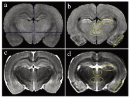
Фигура 1 показывает взвешенные изображения МРТ T1 и T2. По сравнению с контролями, Животные, обработанные пилокарпином, показали высокий сигнал T1 в пироформной коре, боковое таламическое ядро, ретроспециплярный ядро таламуса, и заднее ядро гипоталамуса мозга (Рисунок 1a и b). В T2-взвешенных изображениях животных, обработанных пилокарпином, по сравнению с контролями, Низкий сигнал T2 наблюдался в пироформной коре, соответствует областям высокого сигнала T1 (Рисунок 1C и D). Интенсивности сигналов T2 другого 3 Области высокого сигнала T1 были сопоставимы с областями в контрольной группе (умеренная интенсивность) (Рисунки 1C и D).
Применимость простой в использовании компактной системы МРТ с малым животным в клинической токсикологической патологической исследовании палокарпин. Высокие сигналы T1 и Low T2 показали заметное гистопатологическое повреждение нейронов, Хотя гистопатологическое исследование было более чувствительным.
Использование систем МРТ мелких животных, полученных NIUMAG, имеет большое значение для изучения травмы головного мозга.
 заплесневелый

